



DelphiVet Navigation System: Total Hip Replacement
Professor F. M. Martini
Adopting advanced technology for Total Hip Replacement (THR) might seem like a complex shift, but the reality is that surgical navigation is streamlining workflows and elevating clinical outcomes to unprecedented levels.
During the “Orthopaedic Surgical Navigation: Hands-on Course”, Prof. Martini took the stage to demonstrate the practical applications of the Imaginalis Surgical Navigator in THR. Moving beyond theoretical concepts, he provided a masterclass on how intraoperative navigation bridges the gap between preoperative planning and surgical execution, effectively solving some of the most persistent challenges in veterinary orthopedics.
The Accuracy Gap: Why the Human Eye Isn’t Enough
When it comes to THR, the real challenge in cup and reaming positioning is achieving the perfect angles, specifically, a 45° Angle of Lateral Opening (ALO) and a 20° Angle of Retroversion (ARV). Missing these targets can compromise the entire procedure.
Relying on traditional free-hand techniques leaves a significant margin for error. A 2024 global multicenter study highlighted that only 53.1% of cup placements hit the exact preoperative ALO and ARV targets using standard instrumentation. This unpredictability stems from several factors:
- Dynamic Anatomy: The position of the pelvis frequently shifts during surgery.
- Hidden Landmarks: Clear anatomical landmarks cannot be identified in every case.
- Safety Constraints: Continuous reliance on fluoroscopy raises concerns about X-ray exposure and biosafety.
Surgical navigation changes the game entirely. As Professor Martini demonstrates, the Surgical Navigator provides real-time, millimeter-perfect feedback, turning estimation into a predictable, repeatable standard.
Demystifying the Navigated Workflow
A common hesitation regarding surgical navigation is the fear that it will overcomplicate the operating room routine. Prof. Martini debunks this myth by breaking down the exact workflow, proving how effortlessly the technology integrates into standard practices.
The transition to navigated precision relies on a straightforward, four-step setup:
- Patient Tracker Positioning: Establishing the foundation for continuous, real-time tracking.
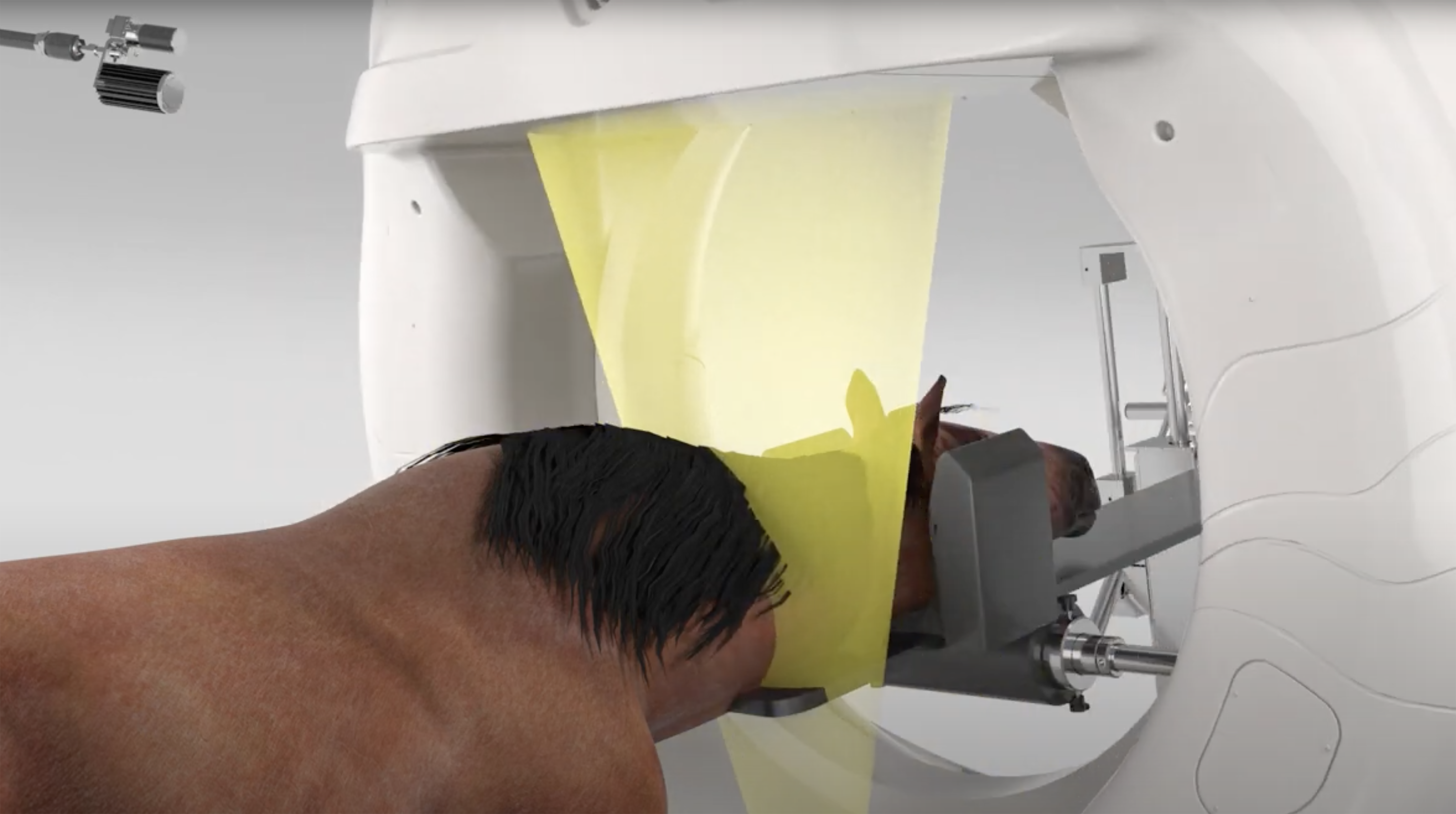
- CT Scan Acquisition: Capturing ultra-accurate 3D anatomical data of the patient.
- Spatial Registration & Surgical Planning: Merging the patient’s physical anatomy with the digital plan with absolute precision.
- Intraoperative Integration: Executing the surgery with real-time video and CT visual guidance.
This seamless integration extends to femur preparation and stem insertion. Dr. Martini highlights the importance of achieving perfect alignment within the femur, ensuring the stem is perfectly centered, with no valgus or retroverted positioning, a task made significantly easier with real-time digital oversight.
Clinical Application: The Complex Case of Dante
To illustrate the true power of surgical navigation, Professor Martini presented the compelling clinical case of Dante, a 9-month-old Maremma Sheepdog. Dante presented with a non-standard hip dysplasia, resulting from a severe post-traumatic pelvic deformity due to an unknown pediatric trauma.
Because of the unique and altered anatomy, standard procedures were insufficient; extreme surgical precision was non-negotiable. The DelphiVet Surgical Navigator provided the real-time, pinpoint accuracy needed to navigate Dante’s complex pelvic structure safely.
For orthopedic surgeons looking to elevate their practice, Dr. Martini’s presentation offers both data-backed insights and exclusive, practical “insider tips” gathered from his extensive hands-on experience. We invite you to watch the full video of Dr. Martini’s speech on our YouTube channel to see the workflow in action, discover the clinical reasoning behind Dante’s surgical plan, and find out which approach the team ultimately chose.
Leave a comment